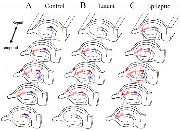
Figure 4. Synaptic Reorganization in mossy fiber pathway results in translamellar hyperexcitability in the hippocampal formation.
The schematic drawings illustrate the normal hippocampal circuitry, the abnormal circuitry during the latent state, and after spontaneous seizures develop in the kainic acid model of mesial temporal lobe epilepsy. The red neurons and axons are excitatory neurons, while the blue neurons are inhibitory interneurons. A. In the normal dentate gyrus, activation of granule cells in a lamella results in a limited activation of the CA3 pyramidal region shown also in red, and the hilar neurons inhibit dentate granule cells in lamellas above and below the activated lamella. B. During the latent state, inhibitory mechanisms are functionally impaired with slow improvement in the inhibitory tone (perhaps, in part, due to synaptic reorganization of inhibitory pathways). There is a mild degree of disinhibition in lamellas above and below the activate lamella (shown as smaller blue block in those lamellas). Furthermore, mild disinhibition results in a greater degree of activation in the CA3 pyramidal region (shown as a greater number of activated red CA3 lamellas). C. Once spontaneous seizures develop in epileptic models, there is prominent synaptic reorganization of the mossy fiber into the inner molecular layer of the DG, across DG blades, and into the basal dendrites of CA3 pyramidal regions. The resulting increased recurrent excitatory connectivity between principal neurons in the hippocampus and within hippocampal lamellas results in translamellar sprouting.

