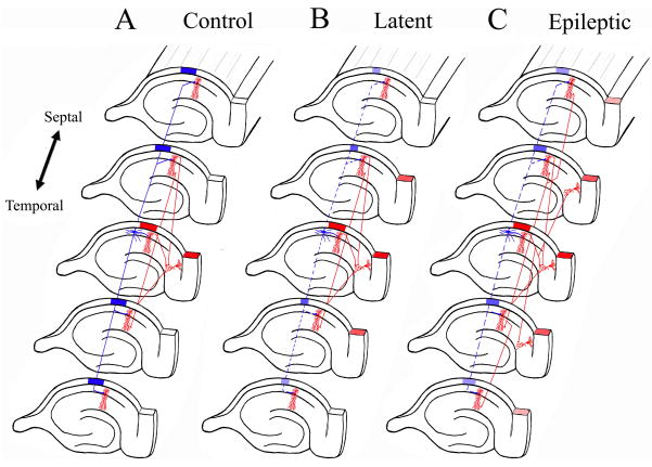
Figure 5. Synaptic Reorganization in CA1 projection to the subiculum results in translamellar hyperexcitability in the hippocampal formation.
The schematic drawings illustrate the normal hippocampal circuitry, the abnormal circuitry during the latent state, and after spontaneous seizures develop in the kainic acid model of mesial temporal lobe epilepsy. The red neurons and axons are excitatory neurons, while the blue neurons are inhibitory interneurons. A. In the normal hippocampus, activation of the CA1 pyramidal neurons in a lamella results in a limited activation of subicular neurons shown also in red, and the CA1 interneurons inhibit CA1 pyramidal neurons from lamellas above and below the activated lamella (shown as a blue block over those lamellas). B. During the latent state, inhibitory mechanisms are functionally impaired with slow improvement in the inhibitory tone (perhaps, in part, due to synaptic reorganization of inhibitory pathways). There is a mild degree of disinhibition in lamellas above and below the activate lamella (shown as smaller blue block in those lamellas). Furthermore, mild disinhibition results in a greater degree of activation in subiculum (shown as a greater number of activated subiculum sections). C. Once spontaneous seizures develop in epileptic models, there is prominent synaptic reorganization of the CA1 pyramidal axons making synaptic contacts with additional CA1 pyramidal neurons and subicular neurons in sections (lamellas) above and below their normal projection pattern. The resulting increased recurrent excitatory connectivity between principal neurons in the hippocampus and within hippocampal lamellas results in translamellar sprouting.

