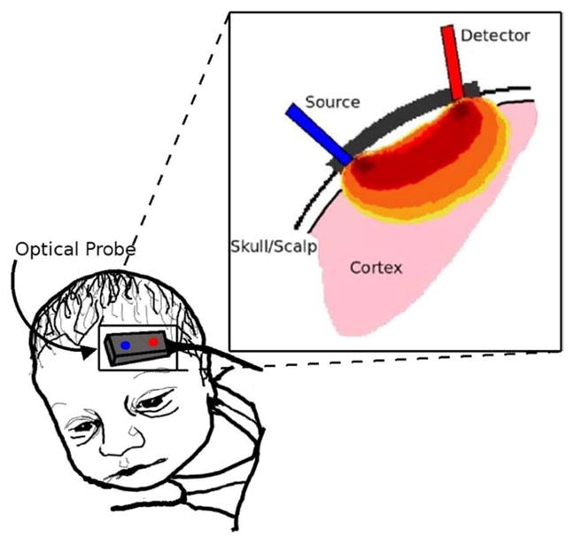
Figure 7.

Depiction of photon propagation through brain tissue. Light that reaches the detector is most likely affected by tissue in the darkest red region, while regions in orange or yellow have less effect on the photons’ journey. The depth of photon penetration is highly dependent on source-detector separation. Larger separations lead to deeper penetration. (Color version of figure is available online.)
