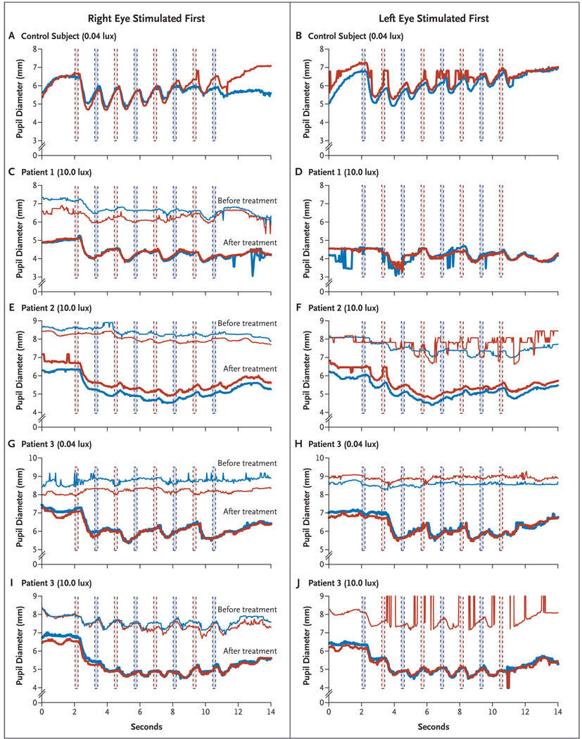
Figure 2. Representative Results of Pupillometry in a Control Subject and before and after Subretinal Injection in the Right Eye of the Three Patients.
Panels A and B show pupillary light reflexes in a control subject after dark adaptation and alternating stimulation with 0.04 lux, starting first in the right eye (red columns) and then in the left eye (blue columns), respectively. The red curves represent the diameter of the right pupil, and the blue curves represent the diameter of the left pupil. The pupillary light reflexes are shown after alternating stimulation with 10.0 lux, starting in the right and then in the left eye, respectively, for Patient 1 at baseline and 4.75 months after injection (Panels C and D) and for Patient 2 at baseline and 2.75 months after injection (Panels E and F). The pupillary light reflexes for Patient 3 at baseline and 1 month after injection are shown after alternating stimulation with 0.04 lux (Panels G and H) and with 10.0 lux (Panels I and J), first in the right eye and then in the left eye, respectively. To facilitate the comparison of overlapping curves in Panel C and Panels E through J, the baseline curves have been shifted up 2 mm with respect to the curves after treatment. The “before” curves were not captured in Panel D or for the left eye in Panel J because of interference from nystagmus.

