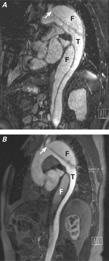
Fig. 7 Multiplanar reformation of A) noncontrast and B) contrast-enhanced magnetic resonance angiography in a patient with type B aortic dissection. Locations of true (T) and false (F) lumina, as well as the proximal entry site (arrow), are seen with both techniques. Of note, a susceptibility artifact due to metallic sternal wires obscures the ascending aorta in the noncontrast image.

An official website of the United States government
Here's how you know
Official websites use .gov
A
.gov website belongs to an official
government organization in the United States.
Secure .gov websites use HTTPS
A lock (
) or https:// means you've safely
connected to the .gov website. Share sensitive
information only on official, secure websites.
