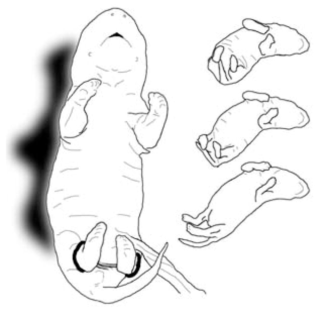
FIGURE 1.

Drawings depicting E20 rat fetuses exposed to interlimb yoke training. The larger drawing on the left, traced from a still photograph of an E20 fetus in a resting posture viewed from a ventral perspective, shows the interlimb yoke attached to both hind limbs at the ankles. The smaller figures on the right present a sequence (top to bottom) of a conjugate limb movement (CLM), in which the two hind legs are extended simultaneously in a caudal direction. These three drawings were traced from individual video frames of an E20 rat fetus recorded after yoke training and removal of the yoke; each image in the sequence is separated from the next by five video frames (0.17 sec).
