
FIGURE 1.
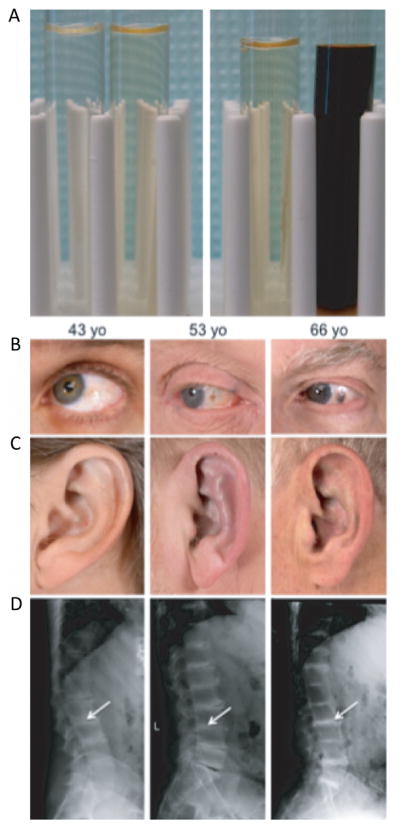
Clinical features of AKU. Detection of excess of homogentisic acid in urine on standing or artificially by adding a base. A: Left panel: Control urine. Left without base added, right with base added. Right panel: AKU urine. Left without base added, right with base added. Three AKU patients with increasing severity (left to right) of clinical symptoms. B: Ochronotic pigmentation of sclerae. C: Auricular cartilages with ochronosis deposition. D: Radiography of dorsolumbar spines: reduction and calcifications of the intervertebral discs (Arrows).