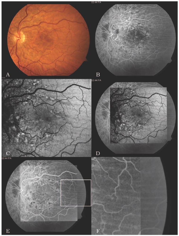
Fig. 1.
A, Color photograph of the left fundus demonstrates predominantly horizontal chorioretinal folds, most prominent in the macula, with subtle peripapillary changes of the retinal pigment epithelium. Findings for the right eye were similar. B, Early-phase fluorescein angiogram of the left eye demonstrates a characteristic pattern of alternating light and dark bands, diagnostic of chorioretinal folds. Linear beadlike areas of hypofluorescence are present around the optic nerve and macula. Findings for the right eye were similar. C, Autofluorescence image of the left eye also demonstrates an alternating pattern of light and dark bands as well as linear beadlike areas of hyperautofluorescence in a distribution similar to that on the angiogram. Findings for the right eye were similar. D, Resulting image after the autofluorescence image (C) is superimposed over the early-phase fluorescein angiogram (B) and aligned in register. E, Resulting image after inverting the autofluorescence image. F, Magnified view of the area in the box in E showing the overlap between the inverted autofluorescence image (on left side) and the fluorescein angiogram (on right side), demonstrating precise alignment but inversion of the alternating banding pattern.

