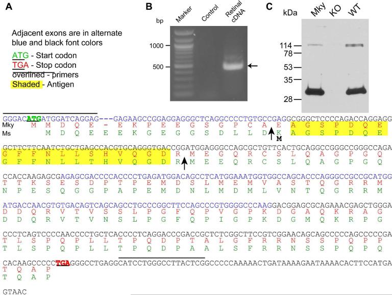
Fig. 1. Retina expresses a new splice variant of PCP2 transcript, Ret-PCP2.
A. The sequence of the PCR reaction product (in blue, now called Ret-PCP2) and its predicted translation in monkey (Mky, red). Mouse (Ms) Ret-PCP2 (green) is shown for reference. The sequences are similar. The arrows mark the translation start point of human cerebellar form-B (first arrow) and form-A (second arrow). Lines above the sequence mark the primers and yellow highlights mark the amino acid sequence used to generate the antibody.
B. A prominent PCR reaction product of ~450 bp (arrow) results from PCR amplification of retinal cDNA. Control includes water instead of cDNA sample.
C. Western blots of monkey (Mky) and wild type mouse (WT) retinal proteins show a single prominent band that migrates with similar mobility (~29 kDa). This band is missing in the PCP2-knockout mouse (KO).

