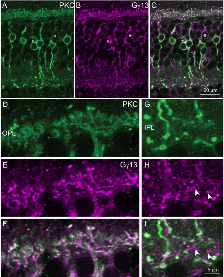
Fig. 10. PKC colocalizes with Gγ13 in the ON bipolar cell dendrites.
Double staining for PKC (green) and Gγ13 (magenta).
A-C. Low magnification shows that some somas are stained only for Gγ13 (*).
D-F. High magnification of OPL shows close to 100% colocalization.
G-I. High magnification of IPL shows that certain bipolar terminals (arrowheads) are stained only for Gγ13. Scale bar in I applies to D-I

