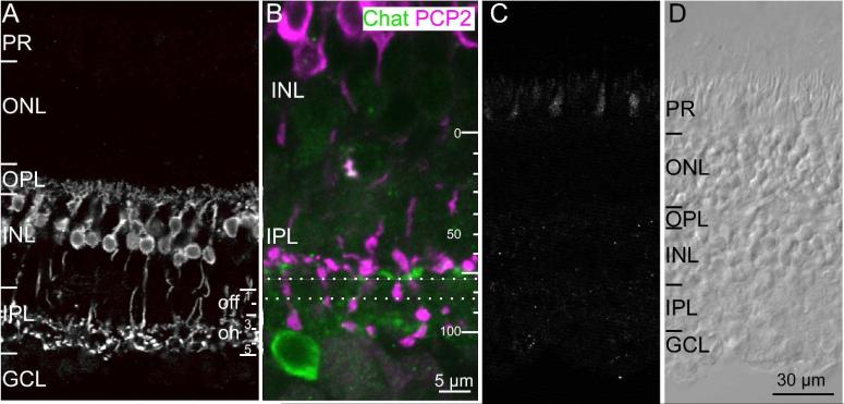
Fig. 2. Ret-PCP2 is expressed by ON cone bipolar cells.
A. Immunostaining for Ret-PCP2 (radial view). Staining is restricted to bipolar cells whose somas are located high in the INL and whose axons terminate in the ON sublamina of the IPL. Note that the arborizations in the IPL appear in two wavy laminas in stratum 3 and 5. For this and all figures: PR, photoreceptors; ONL, outer nuclear layer; OPL, outer plexiform layer; INL, inner nuclear layer; IPL, inner plexiform layer; GCL, ganglion cell layer.
B. Immunostaining for Ret-PCP (magenta) and choline acetyl transferase (ChAT;green) shows that the level of cone bipolar arborization lies just above the stratification of the starburst amacrine cells at around 65% depth. PCP2-stained bipolar terminals hardly arborize between the dotted lines, but axons of rod bipolar cells do cross through this sublamina to arborize in sublamina 5.
C. Preabsorption control. All staining was eliminated in a retinal section that was incubated in antibody that was pre-absorbed with the antigenic peptide (excess of x10). To show some structure, contrast was enhanced relative to the image in B.
D. Image of the same section as in C taken under differential interference contrast.

