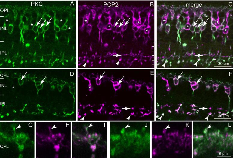
Fig. 3. Ret-PCP2 is expressed throughout diffuse bipolar type 4 (DB4) cells.
Double-labeling for Ret-PCP2 (magenta) and PKC (green).
A-C. Foveal sections.
D-F. Peripheral sections. All PKC-labeled somas are Ret-PCP2-positive (arrows); certain somas are Ret-PCP2-positive but PKC-negative (*). Rod bipolar terminals in sublamina 5 are strongly stained by both antibodies (arrowheads) whereas DB4 terminals are strongly stained for PCP2 but faintly stained for PKC (horizontal short arrows).
G-L. Two examples at higher magnification of dendrites approaching the cones in the OPL: there is a high degree of correlation between the two stains. Arrowheads point to dendrites of rod bipolar cells recognized by their position above the cone bipolar clusters.

