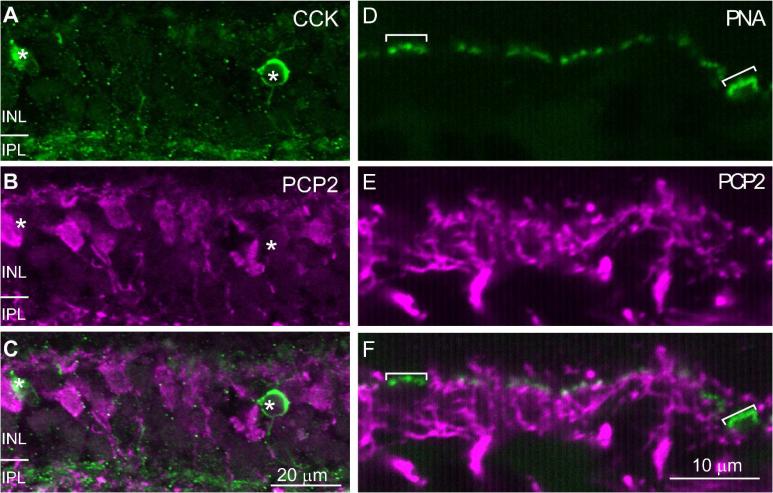
Fig. 5. Ret-PCP2 is not expressed in blue cone bipolar cells.
A-C. Double staining for Ret-PCP2 (magenta) and CCK (green). The images represent a projection of 2-3 confocal images. The two stains did not colocalize in the somas (*). D-F. Double staining using peanut agglutinin (PNA; green) and anti-Ret-PCP2 (magenta). Peanut agglutinin highlights the blue cone pedicles more brightly (brackets) than the other pedicles. These pedicles did not receive Ret-PCP2-stained dendrites.

