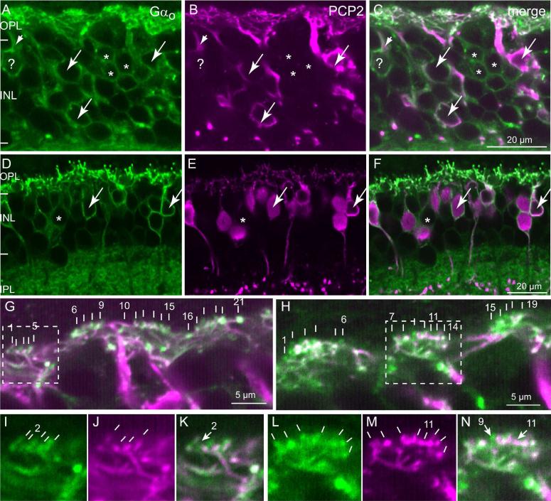
Fig. 7. Ret-PCP2 is localized to the dendritic tips of midget ON bipolar cells.
Double labeling for Ret-PCP2 (magenta) and Gαo (green).
A-F. All Ret-PCP2-positive somas are positive for Gαo (arrows), but certain Gαo-positive somas are devoid of Ret-PCP2 staining (*); Gαo-positive PCP2-negative somas in the fovea (A-C) is much greater than that in the periphery (D-F). Note the Gαo-labeled soma that is marked by ?; it is stained for Ret-PCP2 in its primary dendrite (very short arrow) but not in its soma. Note that the staining for Gαo is restricted to the soma's outline (membrane associated) while that for Ret-PCP2 is in the cytosol.
G–N. High magnification of the outer plexiform layer. G, H are merged images. Each vertical white bar “points” to an ON bipolar invaginating dendrite. Each cluster of dendrites probably resides under a cone pedicle. The first and last dendrites within a cluster are numbered. Dashed squares denote the areas of higher magnification shown in I-N for each stain separately. In I (Gαo), there are 5 resolvable dendrites; only four can be seen in J (Ret-PCP2) (no. 2 is lacking PCP2; arrow in K). In L (Gαo), 7 dendrites are resolvable, while in M (Ret-PCP2) 8 are resolvable. Dendrite no. 11 is strongly stained for Ret-PCP2, but either unstained or weakly stained for Gαo (arrow in N). The slight white color of twig 11 in the merged image (shown by arrow) is due to diffused green background at this spot; the green appearance of twig 9 results from Gαo in this dendrite that extends beyond Ret-PCP2. The orientation of the lines in I-N intends to show the orientation of the dendrites.

