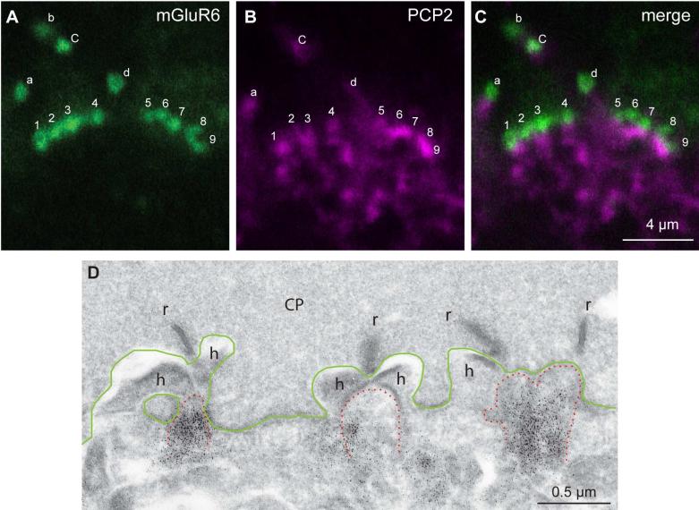
Fig. 9. Ret-PCP2 is expressed in most ON bipolar cell dendrites.
Double staining for Ret-PCP2 (magenta) and mGluR6 (green).
A-C. Two clusters of mGluR6-stained puncta are shown; the lettered puncta are rod bipolar dendritic tips (identified as such because these are large puncta located above the level of the cone pedicles) and the numbered puncta are cone bipolar dendritic tips. mGluR6 concentrates slightly above Ret-PCP2. Rod bipolar magenta punctum b is not visible in this focal plane and d is weak. All cone bipolar puncta can be correlated with Ret-PCP2 staining just below mGluR6.
D. Electron micrograph of monkey retina stained for Ret-PCP2 shows a region of a cone pedicle (CP, its base is outlined in green) with 4 synaptic ribbons (r) and 3 postsynaptic triads. The triads consists of stained (black dots) central elements (ON bipolar dendrites; tip is outlined in dotted red lines) and two lateral elements (horizontal cell process, h).

