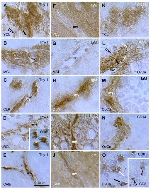
Figure 8.
Staining for Thy-1, IgM, CD8, and CD14, as indicated in panels, in human corpora lutea and ovarian adenocarcinomas (OvCa). YCL, young CL; MCL, mature CL; CLP, CL of pregnancy; RCL, regressing CL (subsequent follicular phase); CAlb, corpus albicans. mv, microvasculature. Scale bar in E applies to panels A-O, including insets. Details in text. Adapted from Ref. [109], © Elsevier.

