
Figure 3.
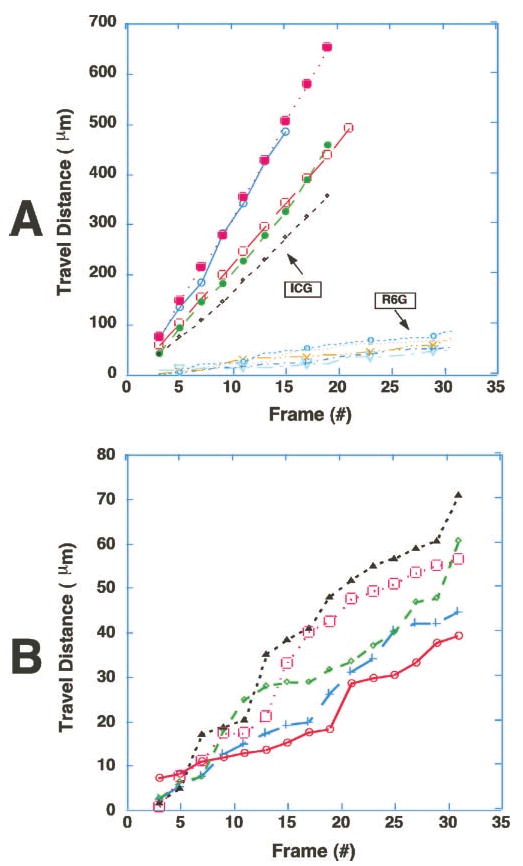
Trafficking patterns of ICG-hyperfluorescent cells were significantly different from those of R6G-stained leukocytes. The time course of spatial positions of five ICG-hyperfluorescent cells and five R6G-stained leukocytes were plotted from video records (30 frame/sec). Note that the ICG-hyperfluorescent cells were moving much faster than R6G-stained leukocytes (A). Furthermore, they were not sticking or rolling on the walls of the blood vessels, in contrast to R6G-stained leukocytes. (B) Expanded plot of the R6G-stained cells in (A), showing characteristic stepwise movement of leukocytes. Video clips of (A) and (B) are available at http://www.iovs.org/cgi/content/full/44/10/4489/DC1.