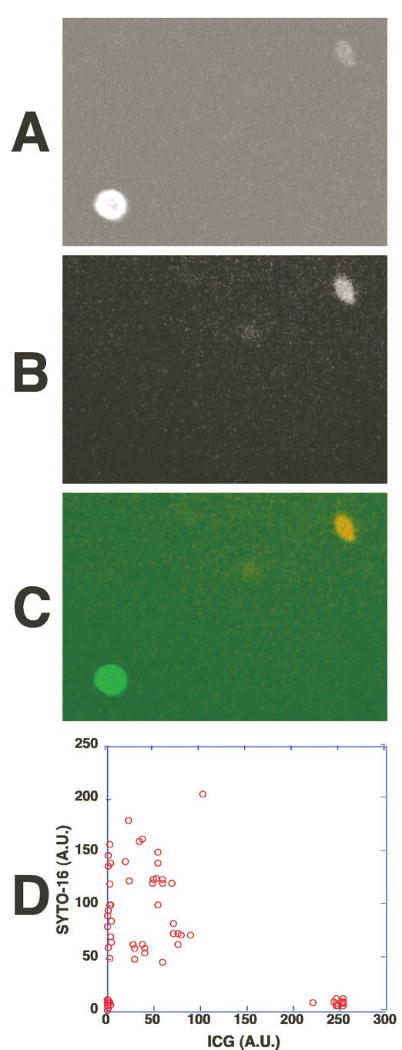
Figure 5.

The strongly ICG-labeled cells did not contain DNA and the weakly ICG-fluorescent cells contained DNA and nuclei. Human whole peripheral blood was incubated with ICG for 1 hour and then stained with a cell-permeating nuclear dye (SYTO-16, Molecular Probes). The fluorescence of ICG and the dye in the cells was examined under the fluorescence microscope. (A) ICG fluorescence of the cells in whole peripheral blood. (B) The DNA staining of the cells shown in (A); 488-nm excitation and 535-nm emission were selected for DNA staining from the nuclear dye's fluorescence. (C) An overlay fluorescence image of ICG (green) and the nuclear dye (red). The strongly ICG-fluorescent cell was SYTO-16 negative, indicating the absence of DNA. A weakly ICG-fluorescent cell contained DNA and a nucleus. (D) The fluorescence intensities of ICG and SYTO-16 fluorescence in each cell were measured and shown in a two-parameter dot plot.
