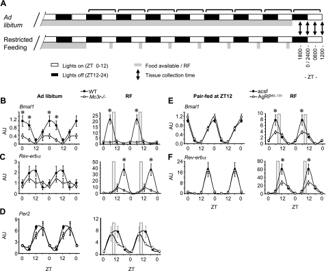
Figure 1.
Mc3r signaling is required for normal liver clock activity during RF. A) Schematic diagram of the RF protocol used for these experiments. Note that food was not available on the day of tissue and serum collection. B–D) Double-plotted expression pattern of Bmal1 (B), Rev-erbα (C), and Per2 (D) in liver of WT and Mc3r−/− mice fed ad libitum or subjected to RF (gray bar). E, F) Double-plotted expression pattern of Bmal1 (E) and Rev-erbα (F) in liver of mice treated i.c.v. with AgRP82–131 or acsf pair-fed a nonlimiting amount of food (4.5 g) at ZT12 (6:00 PM, onset of lights off; left panels) or subjected to RF (right panels). *P < 0.05 vs. corresponding WT. AU, arbitrary units.

