Figure 1.
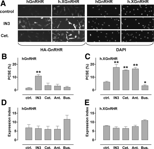
Quantification of HA-GnRHR in HeLa cells by automated imaging. HeLa cells transduced with HA-tagged hGnRHR or h.XGnRHR (1 pfu/nl) were incubated 18 h with IN3, cetrorelix (Cet.), antide (Ant.), or buserelin (Bus.) each at (10−6 m), or no addition (control). They were then stained before image acquisition and analysis as described in the Materials and Methods. Panel A contains representative stains from a proportion (∼10%) of fields showing cell-surface HA-GnRHR or DAPI (nuclear) staining in control and IN3- or cetrorelix-treated cells as indicated. Automated algorithms were used to define cell perimeters, EI, and PCSE values. Panels B and C show PCSE values; panels D and E show whole-cell EI values in arbitrary fluorescence units, each pooled from three experiments with triplicate or quadruplicate wells (mean ± sem; n = 3). *, P < 0.05; **, P < 0.01. ctrl., Control.

