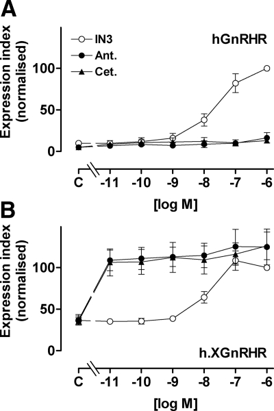
Figure 2.
Concentration dependence of peptide and nonpeptide antagonists effects on GnRHR expression. HeLa cells were treated as in Fig. 1 except that they were incubated for 18 h with 10−11 to 10−6 m IN3, antide (Ant.), or cetrorelix (Cet.) or without antagonist (C, control) before staining for HA-tagged hGnRHR and h.XGnRHR at the cell surface. The data are cell-surface EI normalized a percent of the value obtained for each receptor in the presence of 10−7 m IN3, and are means ± sems (n = 3–7) from seven experiments, each with triplicate wells. The normalized values were significantly greater than control (P < 0.05) for IN3 at 10−8, 10−7, and 10−6 m (at both receptors) and for all concentrations of peptide antagonists at the h.XGnRHR (panel B), whereas the peptides did not increase expression of the hGnRHR at any concentration (P > 0.05, panel A).

