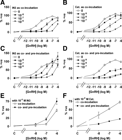
Figure 7.
Influence of IN3 and cetrorelix (Cet.) on GnRH-stimulated NFAT-EFP translocation. HeLa cells transduced with HA-tagged hGnRHR (1 pfu/nl) and the NFAT-EFP reporter were incubated 45 min with 10−11 to 10−6 m GnRH or without agonist (C, control) before imaging. Effects of IN3 (left panels) and cetrorelix (right panels) were also determined by coincubation of agonists and antagonists as indicated (panels A and B). GnRH dose-response curves were also generated in cells preincubated (18 h) with the indicated concentrations of IN3 or cetrorelix and then coincubated (45 min) with GnRH and the same antagonist concentration as used in the pretreatment (panels C and D). The effect of the preincubation is illustrated for a single antagonist concentration by comparison of curves with antagonist coincubation alone, and those with co- and preincubation with IN3 (10−7 m; panel E−data from panels A and C) or cetrorelix (10−8 m, panel F−data from panels B and D). The figures show the proportion of cells in which the N:C NFAT-EFP ratio was more than 1.5, normalized (as a percent of the internal control maximal response). Values shown are means ± sems (n = 3–4) from four separate experiments, each with triplicate wells. GnRH responses with antagonist coincubation differed significantly (P < 0.05) from those with antagonist co- and preincubation at 10−8 to 10−6 m GnRH (for both antagonists, panels E and F). +ve, Positive.

