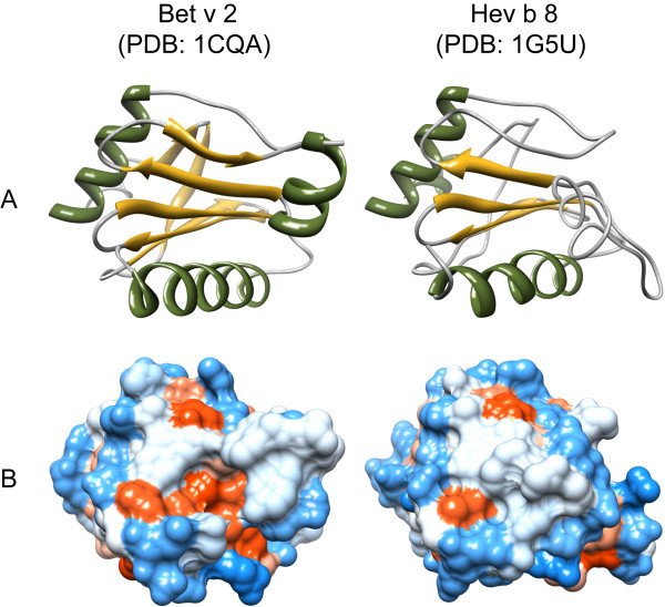
Figure 1.
Three-dimensional structures of allergenic profilins. Secondary structure elements (A) are displayed in green (α-helices) and yellow (β-sheets). The distribution of hydrophilic (blue) and hydrophobic (red) amino acids over the molecular surface is depicted in B. All models were obtained from the Protein Structure Database http://www.pdb.org/pdb/home/home.do and visualized with chimera http://www.cgl.ucsf.edu/chimera/

