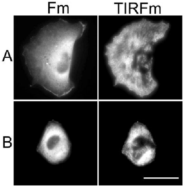
Fig. 3.

CaMK-II is dynamically localized at the surface of motile fibroblasts. Cells were transfected with either GFP-labeled wild-type δC CaMK-II (A) or constitutively active (CON) δC CaMK-II (B) and sub-cultured on FN for 3 h. Cells were imaged using either traditional fluorescence microscopy (Fm) or total internal reflection fluorescence microscopy (TIRFm). Scale bar, 50 μm. Supplemental videos 3 and 4 of live TIRF imaged cells correspond to these conditions.
