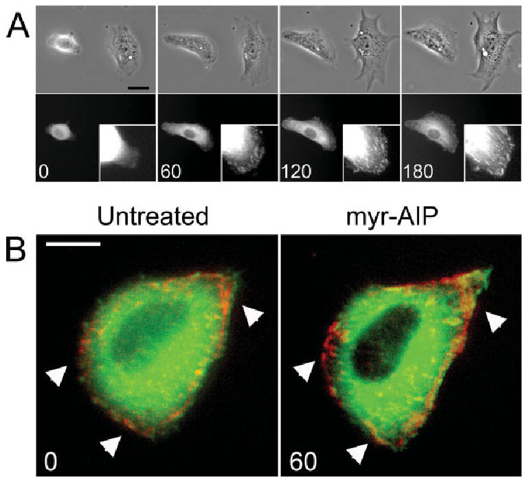
Fig. 6.

Inhibition of CON δC CaMK-II restores paxillin containing adhesions within 1 h. (A) Cells were co-transfected with unlabeled CON δC CaMK-II and GFP-paxillin. Cells were plated on FN for 3 h, re-fed with medium containing 20 μM myr-AIP and then imaged under phase contrast and traditional fluorescence microscopy 0, 60, 120 and 180 min later. Insets represent the lower right-hand corner of the transfected cell. Scale bar, 50 μm. (B) Cells were transfected with CON δC CaMK-II, GFP-vinculin and dsRed-paxillin and then sub-cultured on FN. Cells were then imaged under traditional fluorescent microscopy directly before and 1 h after treatment with 20 μM myr-AIP. Arrowheads indicate regions where myr-AIP treatment induces increased paxillin localization in focal adhesions. Scale bar, 10 μm.
