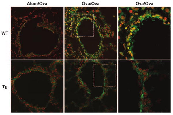
FIGURE 1.

Immunolocalization of NF-κB in airway epithelial cells of allergen sensitized and challenged mice. Frozen lung sections were prepared 48 h following the third daily aerosolized allergen challenge of mock-sensitized (Alum/OVA) or OVA-sensitized (OVA/OVA) mice. Sections were stained with an Ab directed against RelA (Santa Cruz SC-372) followed by incubation with an Alexa 647-conjugated secondary Ab (pseudocolored green). A nuclear counter stain (pseudocolored red) was used to evaluate nuclear localization of RelA, in which case green and red merge to create yellow. Sections were scanned by confocal microscopy. Original magnification of left and center images, ×400. WT, wild-type; Tg, CC10-IκBαsr mice. To better illustrate the differences in nuclear NF-κB localization, the boxed areas of the center panel images were magnified using a ×2.2 optical zoom and are shown on the right. Images are representative of results from six mice per group.
