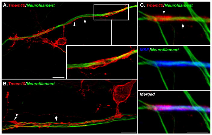
Figure 6. Localization of Tmem10 in myelinated cultures.
A–B. Spinal cord cultures were labeled with an antibody to Tmem10 (red) and neurofilament (green). Inset in A shows a higher magnification of an oligodendrocyte process that is aligned with the axon (arrowhead) and forms a tube that surrounds it. In B, Tmem10 is found along oligodendrocyte processes that are sorting a bundle of axons (arrow). One process ends in a filopodial protrusion (double arrowhead). C. Immunolabeling of myelin segments with antibodies to Tmem10 (red), MBP (blue) and neurofilament (green). Tmem10 is circled around the internodes (arrow) and accumulated at the paranodes (arrowhead). Scale bar: A–B, 10 μm; C, 5 μm.

