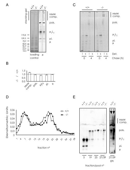
Fig. 4. CHOP promotes optimal assembly of IgM.
A) IgM polymerization was analyzed by non-reducing western blots. Total protein extracts from wt and chop-/- B cells after 4 days of LPS stimulation were subjected to SDS-PAGE in non-reducing conditions and blotted. The nitrocellulose filter was incubated with anti-μ antibodies and the main IgM assembly intermediates were identified. Immuno-decoration with anti-κ or anti-J antibodies (not shown) confirmed the identification of the assembly intermediates.
B) Quantification of the relative amount of each IgM species in chop-/- ASC with respect to the wt. The percentage of IgM species (HMW complexes, polymers, μ2L2, μL, μ) relative to the sum of all the species was calculated after densitometric analyses of non-reducing western blots as the one shown in A. The histogram reports the ratios between each IgM species in chop-/- and wt samples as determined in four independent experiments (n=4; average ± SEM).
C) Wt and chop-/- B splenocytes, stimulated in vitro with LPS (day 3), were pulsed for 10 min with 35S-labeled aminoacids and chased for 0 or 4 hours. NP40 soluble (s) and insoluble (i) fractions were immunoprecipitated with anti-μ and resolved by SDS-PAGE under non-reducing conditions. More HMW complexes are found in the insoluble fraction of chop-/- cells (compare lanes 7 and 3).
D) Lysates from wt and chop-/- B splenocytes, stimulated in vitro with LPS for 4 days, were centrifuged on continuous sucrose gradients. 39 fractions were collected and aliquots analyzed by dot-blot assays with anti-μ antibodies.
E) Individual or pooled fractions of the gradient shown in D, were resolved by SDS-PAGE under non-reducing conditions and blots immuno-decorated with anti-μ to highlight the main assembly intermediates. An enhancement of the two lanes corresponding to fractions 27-39 is shown on the right (Enhanced Pixel Intensity, EPI) to better appreciate the presence of HMW complex. Note that in chop-/- cells HMW complexes (and also some polymers and μ2L2) are more abundant in fractions 27-39 than in control splenocytes.

