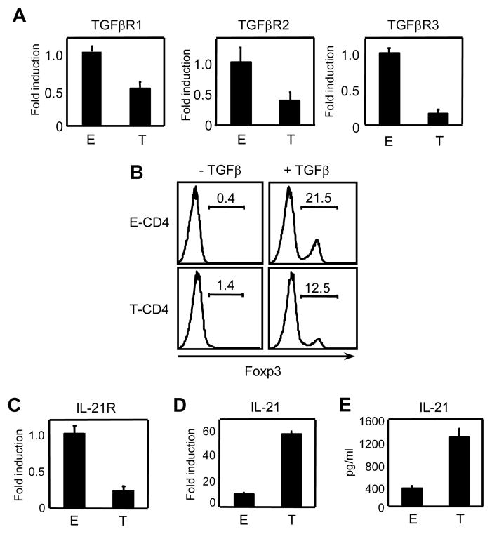
Figure 4. Expression of receptors for TGFβ and IL-21 is reduced in T-CD4 T cells.
(A) RNA was extracted from freshly isolated E- and T-CD4 T cells and TGFβR1, TGFβR2 and TGFβR3 were quantified by qRT-PCR. Fold induction was performed by using the comparative threshold cycle (ΔCT) method and normalized to GAPDH. The shown is the relative expression level in T-CD4 T cells compared that in E-CD4 T cells. (B) Enriched E- and T-CD4 T cells were stimulated with anti-CD3 and CD28 in presence or absence of 5μg/ml of TGFβ for 3 days and analyzed for Foxp3 induction. (C and D) RNA was extracted from freshly isolated E- and T-CD4 T cells and analyzed for IL-21R (C) IL-21 (D) by qRT-PCR. Fold induction was calculated by using the comparative threshold cycle (ΔCT) method and normalized to GAPDH. The shown is the relative expression level in T-CD4 T cells compared that in E-CD4 T cells. (E) Freshly isolated E- and T-CD4 T cells were differentiated under the Th17 inducing condition for 5 days followed by re-stimulation with plate bound anti-CD3. The culture supernatant was analyzed for IL-21 by ELISA. Data shown are representative of at least three independent experiments.

