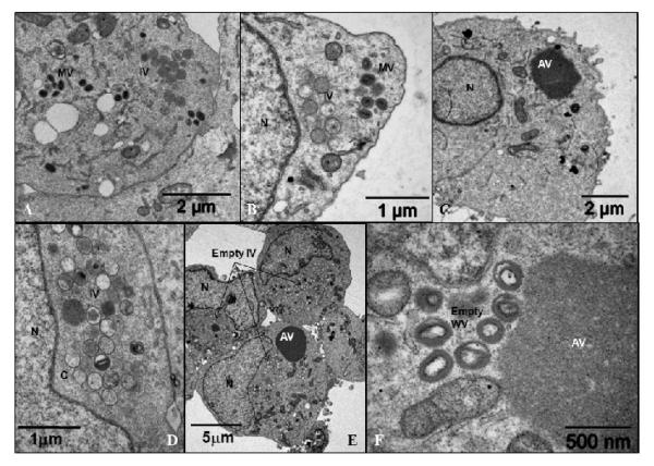
Fig. 5.
Electron micrograph of the vE6i infected cells.
BSC40 cells were infected with vE6i in the presence of IPTG (A, B) or in the absence of the inducer (C – F). After 24 hpi (A – D) or 48 hpi (E – F) the cells were processed for microscopy as described in Methods. IV= immature virions; MV= mature virions; WV= wrapped virions; C= crescents; AV = aggregated virosome; N=nucleus.

