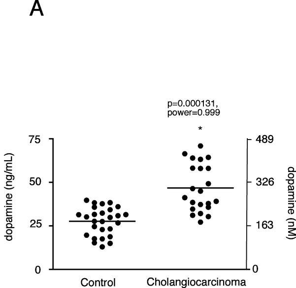
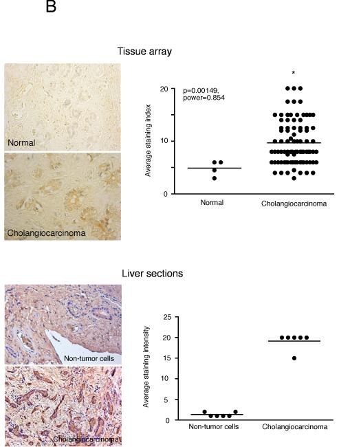
Figure 9.
Dopamine secretion in cholangiocarcinoma. Dopamine levels in bile samples from cholangiocarcinoma patients and patients with intrahepatic cholelithiasis were determined by EIA (A). Data are expressed in a scatter plot of dopamine concentration (ng/mL left axis and nMol right axis). Dopamine immunoreactivity was assessed in biopsy samples from 48 cholangiocarcinoma patients and 4 healthy controls by immunohistochemistry (upper panel) and in paraffin-embedded sections containing cholangiocarcinoma and the surrounding non-malignant liver tissue (n=6; lower panel). Representative photomicrographs of the dopamine immunoreactivity are shown (B; magnification X40). Staining intensity was assessed as described in the methods. P values and the power analysis values are shown in parentheses.


