
Figure 1. Amino acid sequence and features of the expressed BoNT/A derivatives in comparison with wt.
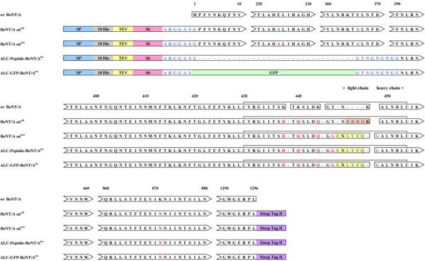
Spaces between arrowheads and arrow tails represent regions of sequence identity omitted for simplicity. Numbers in the upper row correspond to amino acid residues in wt BoNT/A. Residues that are identical in all proteins are shown in black. Introduced mutations are shown in red. Added amino acids are shown in blue. Signal peptide required for insect cell secretion of the expressed derivatives into medium indicated by “SP” on blue background. Tags used for affinity chromatography are indicated: polyhistidine tag is indicated by “10 His” on a gray background; StrepTag II is indicated by “StrepTag II” on a purple background. “TEV” and amino acid sequence on a yellow background represent tobacco etch virus protease recognition sequence. Amino acids on an orange background represent enterokinase recognition sequence. “S6” on a pink background identifies a peptide tag used for site-specific attachment of cargo to the expressed proteins. “GFP” on a green background represents a portion of green fluorescent protein. The five proteins are aligned to illustrate homology between respective structural domains. Gaps have been introduced to facilitate the alignment. Spaces between rectangle-enclosed sequences represent sites of proteolytic cleavage. The disulfide bridges between residues of the light and heavy chains are indicated by horizontal brackets.