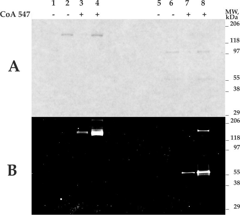
Figure 5. In vitro fluorescent labeling of AcTEV-treated and rEK-treated BoNT/Aadek with Sfp phosphopantetheinyl transferase and CoA 547.
Lanes 1 - 4, unreduced samples; lanes 5 - 8, samples reduced by addition of β-mercaptoethanol. Lanes 1, 3, 5, 7: 0.02 μg BoNT/Aadek; lanes 2, 4, 6, 8: 0.1 μg BoNT/Aadek. Panel A: 10.5 - 14% Criterion gel (Bio-Rad) stained with Bio-Safe Coomassie (Bio-Rad). Panel B: Western blot of gel shown in panel A scanned on a Typhoon 9500 scanner (GE Healthcare) using 300V PMT, 532/580 nm excitation/emission filter set (green).

