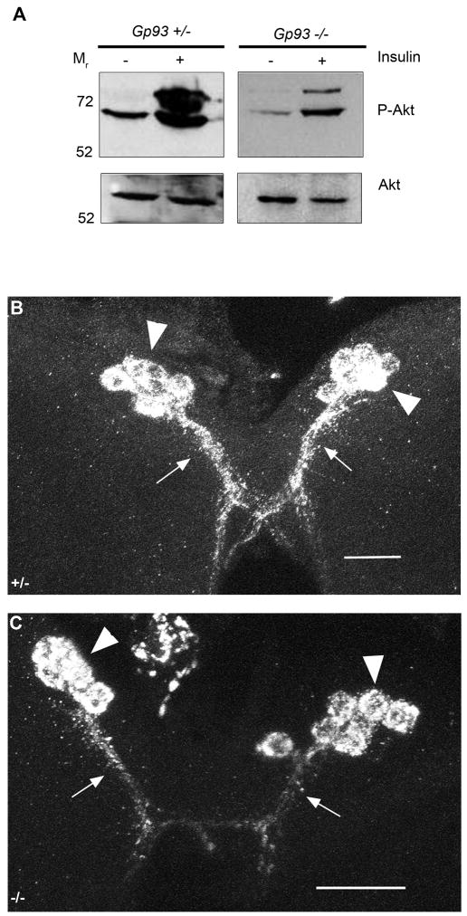
Figure 7. InR function and DILP2 sub cellular localization are unaffected by loss of Gp93 expression.
A) Immunoblot analysis of fat body Akt phosphorylation status in Gp93 heterozygote and mutant larval fat bodies. B–C) DILP2 staining in medial neurosecretory cells in Gp93 heterozygote (B) and Gp93 mutant brain tissue (C). Arrowheads identify cell bodies and arrows the distal axons. Scale bars = 20 μm.

