
Figure 2.
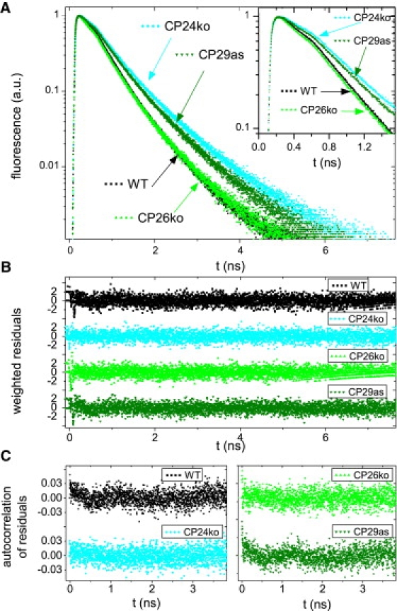
Time resolved fluorescence of thylakoid membranes from WT and mutants of A. thaliana. (A) Normalized fluorescence decay curves. Inset: Initial part of the decay curves. (B) Weighted residuals of the fits of the decay curves in A. (C) Autocorrelation of the weighted residuals. (B and C) Used to evaluate fit quality. The excitation wavelength is 484 nm and the detection wavelength is 680 nm.