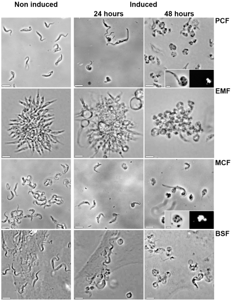
Figure 2. Inducible RNAi on tubulin genes through the T. congolense life cycle.
Phase contrast microscope images of the in vitro cultivated IL3000:13–29 strain transfected with p2T7Ti/αTUB vector in all the developmental stages. Non induced and tetracycline (1 µg/ml for 24 h and 48 h) induced cells are presented. PCF, EMF colonies, MCF (after DE52 purification) and BSF on BAE feeder cell layer were observed directly in the culture medium. In the insets, PCF and MCF were fixed and stained with 4,6-diamino-2-phenylindole (DAPI) before observation in phase contrast. Scale bars = 10 µm.

