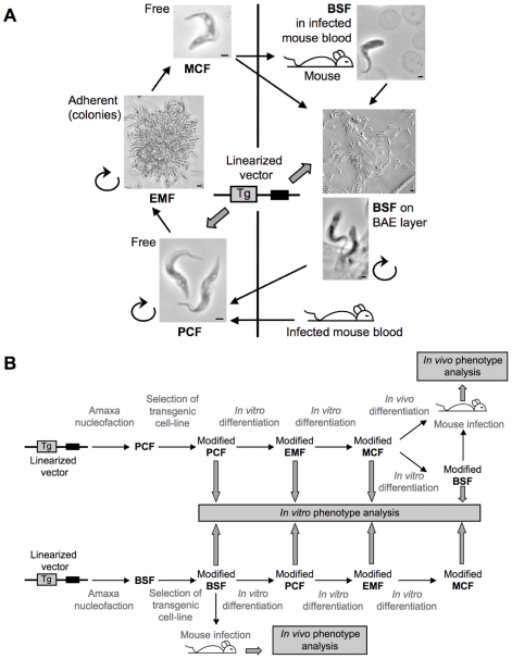
Figure 6. In vitro culture system and genetic tools for T. congolense.
A, The different developmental stages cultured in vitro are represented as individual cells for PCF and MCF as adherent cells forming colonies for EMF and on BAE layer for BSF. Scale bar = 1 mm for PCF, MCF, BSF in infected mouse blood and the lower photo of BSF on BAE layer. PCF, EMF and BSF are dividing cells as represented by the rounded arrow. BSF differentiation can be achieved either by infection of mice and culture from blood or directly in vitro on BAE layer. B, Scheme of transgenic cell-line analysis through the cycle starting from PCF (top) or BSF (bottom) transfection. Linearized vectors used in transfection assays are represented by a line and two boxes, the black one represents the selection marker and the grey one represents the transgene (Tg).

