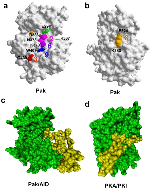
Figure 7. The protein surface representation of Pak2 and PKA, identification of reciprocal coupling pairs, and association with inhibitory peptides.
(a) Identification of the Pak2 reciprocal coupling residues that are on the Pak1 surface (1YHV.PDB). (b) The structure turned 120 degrees counterclockwise from (a) shows the reciprocal coupled ion pair in the hinge region. (c) The catalytic domain of Pak (green) binding to the AID (yellow) modified from 1F3M.PDB. (d) PKA (green) bound to PKI (yellow) modified from 1ATP.PDB.

