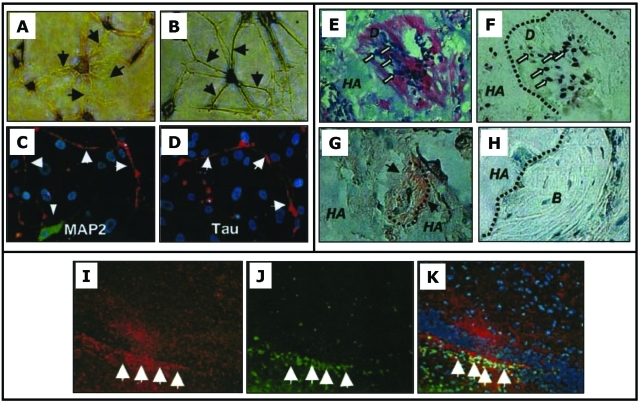
Figure 2.
In vitro neurogenesis of SHED (A-D), transplanted SHED into immunocompromised mice (E-H) and into the mouse brain (I-K). (A,B) Toluidine blue staining of the altered morphology of SHED after neurogenic induction. (C,D) Immunopositive staining for MAP2 and Tau on dendrites and axons (arrows), respectively. (E,F) Eight wks after transplantation into the subcutaneous space, SHED differentiate into odontoblasts (open arrows) and form dentin-like structure (D) on the surfaces of HA. The same field is shown for human-specific alu in situ hybridization, indicating the human origin of odontoblasts (open arrows in F). (G) Immunohistochemical staining of DSPP on the regenerated dentin (black arrows). (H) Newly generated bone (B) by host cells in the same SHED transplant shows no reactivity to the DSPP antibody. (I-K) Neurogenically induced SHED injected into the dentate gyrus of the hippocampus of immunocompromised mice for 10 days. (I) NFM (red) and (J) human-specific anti-mitochondrial antibody (green) and (K) merged images showing co-localization of the two (adapted from Miura et al., 2003, with permission).

