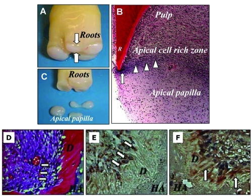
Figure 3.
The anatomy of the human apical papilla (A-C) and dentinogenesis of SCAP in immunocompromised mice (D-F). (A) An extracted human third molar depicting root attached to the root apical papilla (open arrows) at the developmental stage. (B) Hematoxylin and eosin staining of human developing root (R) depicting epithelial diaphragm (open arrows) and apical cell-rich zone (open arrowheads). (C) Harvested root apical papilla for stem cell isolation. (D) Eight wks after transplantation, SCAP differentiated into odontoblasts (arrows) that formed dentin (D) on the surfaces of a HA carrier. (E) SCAP differentiated into odontoblasts (arrows) are positive for anti-human specific mitochondria antibody staining. (F) Immunohistochemical staining of SCAP-generated dentin (D) showing positive anti-DSP antibody staining (arrows) (adapted from Sonoyama et al., 2006, 2008, with permission).

