
Figure 2.
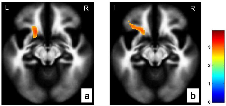
The axial-oblique images (MNI coordinates z=-12mm) display the regions in ventrofrontal white matter in which fractional anisotropy showed a positive association with functional coupling from perigenual anterior cingulate cortex to amygdala during (a) fearful processing (MNI coordinates for the point of maximal association: x= -18mm, y= 18mm, z= -10mm, 107 voxels, T=3.32, P=0.001 uncorrected) and (b) happy processing (MNI coordinates for the point of maximal association: x= -18mm, y= 20mm, z= -10mm, 157 voxels, T=2.88, P=0.002 uncorrected) in the entire cohort. The color bar represents the range of T values. The findings are displayed on a tissue probability map of white matter. L: left brain; R: right brain.