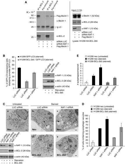
Figure 5.
BCL-2 inhibition of Beclin 1-dependent autophagy requires NAF-1. (A) NAF-1 contributes to the interaction between BCL-2 and Beclin 1. H1299 HA-BCL-2b5 cells were transfected with Flag-Beclin 1 and either LUC or NAF-1 siRNA. Cells were lysed and subjected to immunoprecipitation with anti-BCL-2 antibody. Precipitates were subjected to analysis by immunoblot using anti-Beclin 1 and anti-BCL-2. All lanes are derived from the same gel and of the same exposure. Thin white lines indicate where lanes have been removed. (B) Loss of NAF-1 prevents BCL-2b5 from antagonizing starvation-induced autophagy. H1299 GFP-LC3 and BCL-2b5/GFP-LC3 cells were transfected with either LUC or NAF-1 siRNA and starved for 4 h. Cells were analysed as in Figure 4C. (C) Representative electron micrographs of H1299 neo and HA-BCL-2b5 cells treated with either LUC or NAF-1 siRNA, with or without subsequent starvation. Scale bar represents 10 μm. Cell lysates were analysed by immunoblot. All lanes are derived from the same gel and of the same exposure. Thin white line indicates where lanes have been removed. Autophagy levels observed by electron microscopy (C) were quantified and expressed as either the percentage of cells containing autophagic vacuoles (D) or the number of autophagosomes per cell (E). A minimum of 100 cells per sample were counted; results represent the average±s.e.m.

