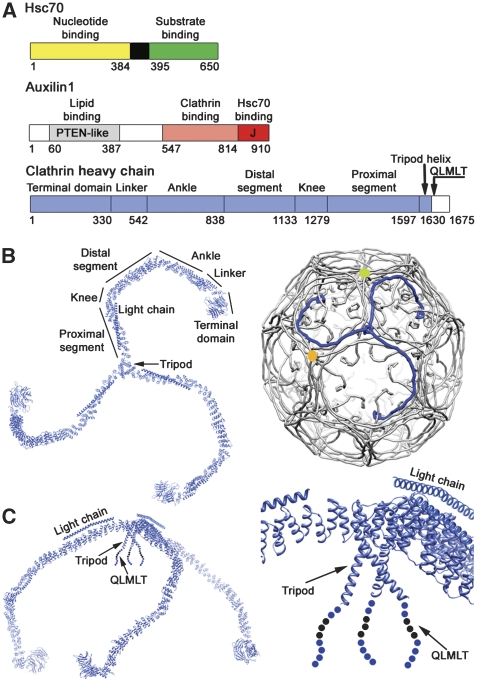
Figure 1.
Components of the clathrin uncoating process. (A) Domain organization of Hsc70 (top), auxilin (middle) and clathrin heavy chain (bottom). Residue numbers for domain or regional boundaries are shown below the bars. (B) A clathrin triskelion (left) and its packing within the lattice of a coat (right). The various regions of the heavy chain are labelled; the ordered, 71-residue α-helical segment of the light chain is also shown. Three symmetry-distinct vertices are colour-coded, yellow, blue (the hub of the blue triskelion) and green. (C) Side view of the triskelion (left), illustrating the pucker at the apex, and a close-up of the hub region, including the helical tripod and the QLMLT sequence near the C-terminus.

