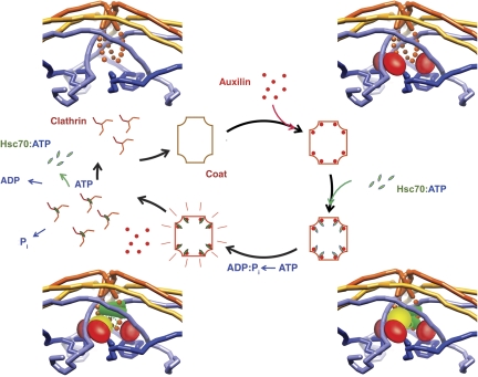
Figure 7.
Model for the uncoating mechanism. The central diagram is a schematic representation of the underlying Hsc70/clathrin cycle, and the four corner diagrams show details of binding events at a vertex. Clockwise, from upper left: clathrin coat binds auxilin (red), which stabilizes a strained clathrin conformation (manifested by change in axial ratio of coat); auxilin recruits Hsc70:ATP (ATPase domain in yellow; substrate-binding domain in green); Hsc70 cleaves ATP and substrate-binding domain clamps tightly onto a specific segment of the disordered C-terminal tail of the heavy chain, trapping further strain in the clathrin lattice; when a large enough number of vertices have bound Hsc70, the accumulated strain causes the coat to dissociate, releasing auxilin, clathrin:Hsc70:ADP and Pi. Nucleotide exchange and dissociation of Hsc70 from clathrin complete the cycle.

