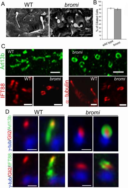
Figure 5. Cilia defects in bromi mutants.
(A) Neuroepithelial cilia from E10.5 wild-type (WT) and bromi mutant neural tubes analyzed by scanning electron microscopy. bromi mutant cilia showed a swollen or bulbous morphology (arrowheads). (B) Cilia frequency in the wild-type and bromi mutant neuroepithelium. (C) Confocal images of neural tube cilia stained with Arl13b (upper panels), IFT88 and acetylated α-tubulin (lower panels). (D) Neural tube cilia from E10.5 wild-type (WT) and bromi mutant embryos immunostained for γ-tubulin/Arl13b/ Gli2 or γ-tubulin/IFT88/Gli2. Scale bars are 1 μm in A and C and 0.5 μm in D. Error bars in B reflect +/− standard error of the mean.

