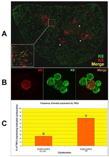
Figure 2.
K8+K5+ cells exist at the cortico-medullary junction and are a subset of TNCs. Confocal analysis of keratin and pH91 staining of thymic sections and freshly isolated TNCs. (A) Thymic section stained with K5 (red) and K8 (green). Yellow regions (inset and asterisks) show double positive (K8+K5+) epithelial cells. Original magnification: 40X. (B) Freshly isolated TNCs stained with K5 (red) and K8 (green). The double positive TNC appears as yellow in the merge. Original magnification: 40X. (C) Frequency of K8+ and K8+K5+ populations in freshly isolated TNCs determined by manual counting of 1000 cytospun TNCs after immunostaining. No K5+ single positive cells with the TNC complex morphology were detected. Data for (A) and (B) is representative of three independent experiments.

