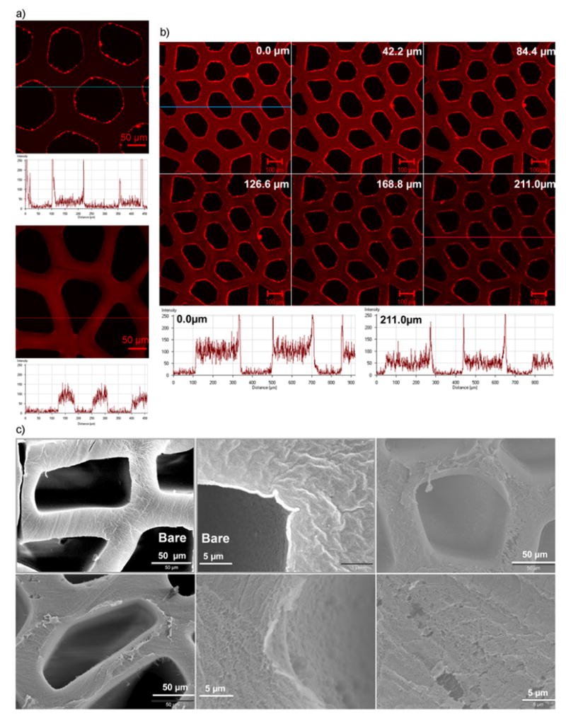
Figure 2.

Confocal and scanning electron microscopy images showing the formation of LbL assembled multilayer thin films on agarose hydrogel substrate. a) Confocal microscopy images and associated fluorescence intensity profiles of a section below the top surface of agarose scaffolds coated with 30 bilayers of PAA and PEG followed by three bilayers of PEG and TRITC conjugated BSA (top image), and of agarose scaffolds coated with only 3 bilayers by PEG and TRITC conjugated BSA (bottom image). b) Confocal microscopy acquired z-section images of agarose scaffolds coated with 30 bilayers of PAA and PEG followed by three bilayers of PEG and TRITC conjugated BSA. Fluorescence intensity profiles of the topmost section (top panel: leftmost image) and a middle section (bottom panel: rightmost image) are shown. c) Scanning electron microscopy images of the scaffolds LbL coated with 30 bilayers of PAA and PEG. “Bare” denotes the non-coated scaffolds. BPEI was used as the LbL initiating polyelectrolyte for PAA/PEG assemblies.
