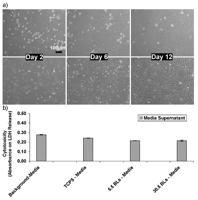
Figure 8.

a) Phase contrast microscopy images demonstrating the cytophobicity of the 30.5 bilayers of PAA/PEG multilayers over time (days). Top panel: NIH-3T3 fibroblasts cells on TCPS plates coated with 30.5 bilayers of PAA/PEG. Bottom panel: Fibroblasts on bare TCPS plates (control). b) Cytotoxicity levels of fibroblast cells after 4 days of culturing on (PAA/PEG)5PAA multilayers built onto TCPS (denoted here as 5.5BLs), (PAA/PEG)30PAA multilayers built onto TCPS (denoted here as 30.5 BLs), and bare TCPS plates. Serum in the cell culture medium contains a small amount of LDH, which is shown as “background media”. The absorbance values corresponds to the amount of lactate dehydrogenase (LDH) released into the culture supernatant.
