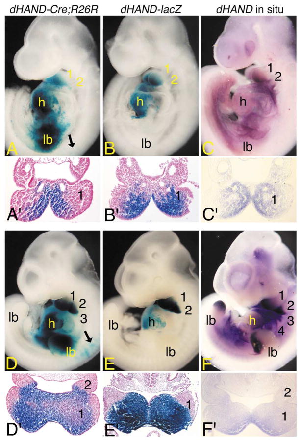
Fig. 2.
Early dHAND expression domains in dHAND-Cre; R26R embryos. (A–F) Lateral (A–F) and transverse (A′–F′) views of dHAND-Cre;R26R (A, A′; D, D′) and dHAND-lacZ (B, B′; E, E′) embryos stained in whole mount for β-gal activity and wild type embryos (C, C′; F, F′) after whole-mount in situ hybridization analysis of dHAND expression. Transverse sections of β-gal-stained embryos were counterstained with nuclear fast red and photographed by using brightfield optics. Transverse sections of whole-mount in situ embryos were photographed by using Nomarski DIC optics. (A) In E9.5 dHAND-Cre; R26R embryos, labeled cells are observed in the first mandibular (1) and second (2) arches, common heart ventricle (h), and forelimb bud (lb). Few scattered cells are observed in the early dorsal root ganglion (black arrow). (A′) A transverse section through the first arch of the embryo shown in (A) illustrates that stained cells are confined to the arch mesenchyme in a dHAND-specific pattern. (B) In E9.5 dHAND-lacZ embryos, staining is observed in the mandibular arch and common heart ventricle. (B′) A transverse section through the first arch of the embryo shown in (B) shows that labeled cells are confined to the arch mesenchyme. (C) In E9.5 wild type embryos, dHAND expression is observed in the first mandibular and second arches and in the future right ventricle of the heart (obscured by the left ventricle). (C′) As observed in (A′) and (B′), dHAND expression is confined to the arch mesenchyme. (D) By E10.5, labeled cells in dHAND-Cre;R26R embryos are observed in arches 1–3, the ventricle, and the limb bud. Scattered cells are also observed in the atrium. While not visible in this view, stained cells are also observed in arch 4. Labeled cells are also observed in the cervical dorsal root ganglion (black arrows). (D′) In a transverse section through the first and second arches, staining is observed in the arch mesenchyme, with the intensity of staining highest in the periphery of the arch. (E) In E10.5 dHAND-lacZ embryos, labeled cells are observed in arches one and two and in the ventricles of the heart. Scattered cells are also present in the atria. (E′) As observed in dHAND-Cre;R26R embryos, staining within the mesenchyme of the first arch is strongest at the periphery. While not shown due to a slight difference in the angle of section, the second arch staining in dHAND-lacZ embryos is comparable with that of dHAND-Cre;R26R embryos. (F) dHAND expression in E10.5 wild type embryos is observed in arches 1– 4, distal limb buds, and right ventricle of the heart. (F′) A section through the first and second arch again illustrates staining throughout the medial half of the arch mesenchyme, with the strongest staining observed along the periphery of the arch.

