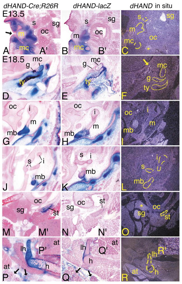
Fig. 4.
Distribution of dHAND progeny cells in the middle ear and throat during development of dHAND-Cre; R26R embryos. Transverse (A–C; E13.5) and frontal (D–R; E18.5) sections through the middle ear and throat of dHAND-Cre;R26R (A, D, G, J, M, P), dHAND-lacZ (B, E, H, K, N, Q), and wild type (C, F, I, L, O, R) embryos. Treatment of sections was performed as described in Fig. 3. (A–C) Sections through the middle ear of E13.5 embryos. (A) In dHAND-Cre;R26R embryos, stained cells are observed in the mesenchyme surrounding the epithelium of the external auditory meatus (black arrow), in Meckel’s cartilage (mc), and in the future manubrium of the malleus (m). Scattered labeled cells are also observed in the sympathetic ganglion (sg; see inset, A′). Labeled cells are not observed in the stapes (s), though are present in more proximal sections (see text). (B) Labeled cells in dHAND-lacZ embryos are also observed in the mesenchyme of the middle ear region, the manubrium of the malleus, and Meckel’s cartilage, though they are mixed with unlabeled cells in Meckel’s cartilage. No staining is observed in the stapes or sympathetic ganglion (see inset, B′). (C) Very little endogenous dHAND expression is observed in the middle ear. The exceptions are the sympathetic ganglion and the mandibular branch of the trigeminal ganglion (*). (D–R) Frontal sections through the heads of E18.5 embryos. (D) The tympanic (ty) and gonial (g) bones and Meckel’s cartilage are composed almost solely of labeled cells in dHAND-Cre;R26R embryos. The surrounding mesenchyme in this region of the head is also composed of labeled cells. (E) Labeled cells in dHAND-lacZ embryos are observed in the tympanic ring and gonial bone, though a small portion of unlabeled cells are also present. More significant mixing of labeled and unlabeled cells is observed in Meckel’s cartilage. (F) Little endogenous expression is observed in the tympanic and gonial bones and Meckel’s cartilage, though faint expression is found along some of the head vasculature (arrow). (G, H) In sections through the malleus, labeled cells in dHAND-Cre;R26R and dHAND-lacZ embryos are confined to the manubrium (mb) of the malleus and surrounding mesenchyme. (I) dHAND expression is not observed in any part of the malleus or incus (i) in wild type embryos. (J, K) β-gal-stained cells in dHAND-Cre;R26R and dHAND-lacZ embryos are not present in the incus or stapes (s). Labeled cells in the manubrium of dHAND-lacZ embryos are mixed with unstained cells. (L) dHAND expression is not observed in the incus, stapes, or manubrium of the malleus. (M) In sections through the distal portion of the styloid cartilage (st), labeled cells in dHAND-Cre;R26R embryos compose most of the styloid and are scattered in the sympathetic ganglion (see inset, M′). (N) In dHAND-lacZ embryos, labeled cells are present in the perichondrium of the styloid cartilage, but are mixed in the cartilage itself and are absent in the sympathetic ganglion (see inset, N′). (O) dHAND expression is observed in the sympathetic ganglion in the incus/stapes region, with weaker expression in the glossopharyngeal ganglion. (P) In the throat, β-gal-stained cells in dHAND-Cre;R26R embryos are observed in the lesser horn (lh) and body (h) of the hyoid. However, labeled cells are mixed with unlabeled cells in the body, possibly reflecting changes in staining in cartilage undergoing endochondral ossification, as few hypertrophic chondrocytes are stained. Labeled cells are also observed in the mesenchyme of the submandibular and sublingual glands (black arrows). Out of the photographic frame, adipose tissue also contains scattered labeled cells (inset, P′). (Q) As with other cartilages, labeled cells in dHAND-lacZ embryos compose the perichondrium of the lessor horns of hyoid but are mixed with a majority of unlabeled cells in the cartilage itself. Very few labeled cells are observed in the body of the hyoid. Labeled cells are observed in the mesenchyme of the submandibular and sublingual glands (arrows), but are absent in adipose tissue observed out of the photographic frame (inset, Q′). (R) dHAND expression is not observed in or around the hyoid cartilages or bones; however, adipose tissue at the base of the neck shows strong dHAND expression. Out of the plane of section, expression is also observed in the submandibular gland (inset, R′). oc, otic capsule.

